Pancee Dental Clinic Pondicherry secured the Orthophos SL 3D Imaging Machine, catapulting us onto the Digital Dentistry platform with a firm foundation in imaging technology. ORTHOPHOS SL’s intuitive user interface and automatic positioner aids in precise image acquisition, minimizes waiting times, avoids the need for corrections while and guaranteeing perfect results.
A new dimension in diagnosis.
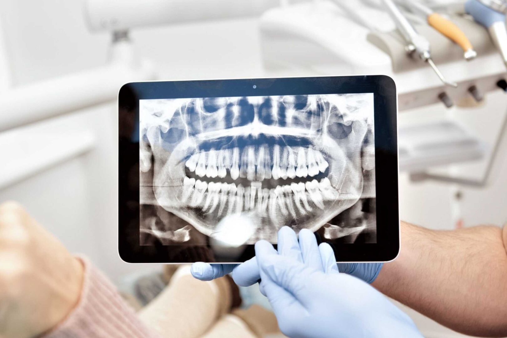
Healthcare technology is evolving for the betterness of the mankind day by day, especially imaging technology. Kamala Dental is trying to keep dental imaging and diagnosis instant and accurate, thus we incorporated the CBCT into our practice. 3D evaluation of oral and facial structures using CBCT helps in safer, better, faster and an accurate diagnosis. Kamala Dental introduced the Sirona Orthophos SL 3D from Germany, the latest of its kind to serve you better.
CBCT gives a better imaging with accurate measurements in a various views and angles, which make the evaluation, diagnosis and treatment planning foolproof. The radiation dose used with a CBCT scan is much less compared to that of a regular CT scan. It’s so quick that the scan normally takes between 20-40 seconds for a full mouth scanning and less than 10 seconds for a scan of a specific area. This makes the evaluation and diagnosis of your dental problems at Kamala Dental possible in the very first visit.
Other than diagnosis, CBCT is a great tool for treatment planning, and in execution, that makes Pancee Dental Clinic Pondicherry purely, digital. With its low cost, low radiation and the ease of use, the Kamala Dental team uses this as a routine imaging technique for all dental treatments. Along with intraoral scanning, which provides a soft tissue profile and CBCT that generates a bony architecture, and these both images can be merged to create a virtual patient for the better diagnosis, treatment planning and execution, that too with low radiation values and lesser expense – it’s a revolution in dental diagnosis and treatment.
Implantology
Dental Implants placed in rehabilitation of missing teeth, exactly on the long axis of the future crowns, provides a better anchorage and better bone support in the long term. Such placements are done without injuring the vital structures such as the nerves, blood vessels, and sinus membranes, can only be assured by planning three dimensionally using CBCT imaging.
Single, multiple teeth or full mouth rehabilitations, well-planned digital protocols that are executed using a surgical guide are the latest treatment protocols at Pancee Dental Clinic Pondicherry.
Endodontics/ Root canal treatments
The root canal system in our teeth, is really complex and shows huge variations in number and shape from person to person. It’s interconnections and divisions are not possible to be evaluated two dimensionally. At Pancee Dental Clinic Pondicherry, root canal treatments are no more a guesswork.
Knowing the number of the root canals and its varying complexities in advance, will assist the endodontists at Pancee Dental Clinic Pondicherry, to provide you the best results through their hands. The reason for a failed root canal treatment can be best assessed using this three-dimensional diagnostic method (CBCT), and the offending (painful) tooth can be salvaged.
Surgeries
Minor and major surgeries in the maxillofacial region and the reason for the pain is either dental related or due to lesions of the jaw. These lesions can be best investigated with the three-dimensional scan (CBCT). The extent of the lesion and the involvement of vital structures are accurately evaluated and the surgical correction is done with minimal invasion and trauma as well as high precision.
Impacted teeth Removal
Impacted teeth removal is one of the most common procedures done in our dental practice. Via this procedure, the removal of the locked-in and infected wisdom teeth, supernumerary teeth, and at times a few front teeth are done. The surgery is performed in a planned manner whilst selecting the part for removal and tooth division if necessary, thus assuring minimal opening for the surgery. The three-dimensional evaluation of such impacted teeth and its proximity to vital structures such as the inferior alveolar nerve, is analyzed using CBCT and these cross-sectional images will be used for planning the surgery, minimizing temporary or permanent damage to any of these structures during the surgical procedures.